
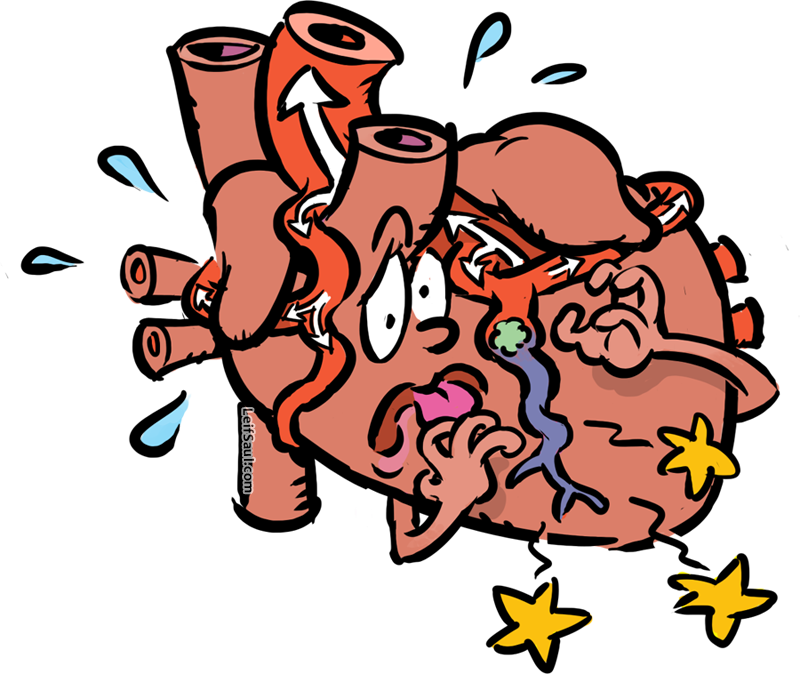
This week’s image may seem melodramatic, but I’m sure if you were a heart undergoing a heart attack, you’d feel something like this.
Heart attacks teach us that the heart is like any other organ – it consists of cells that require oxygen and nutrients to survive. The other thing we learn is that, despite the large amount of blood in the heart, almost none of its contents are directly available to the heart’s cells. That’s because diffusion is only effective across short distances, so the thickness of the heart’s walls prevents all but the inner surface cells (endothelium) from acquiring their vital resources in this way.
The solution is for the heart to have its own network of blood vessels, the coronary arteries, penetrating deep into the heart walls to supply its own tissues with blood. The suffering heart depicted here is actually anatomically correct (minus the face and heads), allowing us to see all the major blood vessels that deliver blood to and from the body.
Oxygenated blood, from the lungs, reaches the heart through the four pulmonary veins, entering horizontally from the sides. It enters the left side of the heart, which ejects this oxygen-rich blood (shown in red) into the aorta, the largest artery in the body. The aorta emerges diagonally from the left side and arches over the top of the heart, branching many times to provide oxygen to the entire body.
From the aorta, the right coronary artery curves around the heart’s right side (on the left side of the page), and the left coronary artery curves toward the heart’s left side, each providing many branches of their own. If one of these becomes blocked by a blood clot or fatty deposits in the artery walls, then a heart attack results – meaning the heart tissue dies due to the obstructed blood supply, and the resulting lack of oxygen to that region. This is different from cardiac arrest, in which the heart stops beating – a problem with the electrical conducting system of the heart. (However, the two problems can be related, if a heart attack damages part of the conducting system.)
Let’s look at the rest of the blood flow, in a healthy heart. From a coronary artery, tiny branches (capillaries) supply oxygen to the heart’s tissues by diffusion, and drain into cardiac veins (not shown) which drain into the right side of the heart. This is also where oxygen-depleted blood enters from the rest of the body, through the superior and inferior vena cava (vertical vessels on the left side of the page). From there, it is pumped through the pulmonary trunk (large vessel crossing in front of the aorta) back to the lungs.